
Elbow Dysplasia or Elbow Displeasure?
- Dr. Darryl Millis

- Feb 29, 2020
- 7 min read
Undeniably, elbow dysplasia leads to elbow displeasure. Recently, we polled our My Lame Dog Facebook group regarding the likelihood of a 10-11 month old dog with a fragmented coronoid process (part of the elbow dysplasia complex that results in a fragment from the weight bearing portion of the ulna in the elbow joint) having end stage (i.e. bone on bone) osteoarthritis. It is always amazing to me how quickly dogs can develop severe cartilage damage as a result of elbow dysplasia.
Most of the general public associates end stage osteoarthritis with old age. Uncle Joe with a total knee at age 68, Aunt Sadie with a total hip at the age of 75, and so on. Yes, dogs can get wear and tear arthritis associated with aging. But most of the severe cases of arthritis in young dogs comes from abnormal formation of joints, such as elbow dysplasia or hip dysplasia. Literally, dysplasia means abnormal formation and development of a joint. Arthritis may result from abnormal forces on normal joints, or normal forces on abnormal joints. Elbow dysplasia is the result of normal forces on abnormal joints. As such, the development of arthritis can occur very rapidly, and to a severe extent. The elbow is a complex joint, with the humerus, ulna and radius all needing to fit together like a glove. If one bone is a bit longer or shorter than the other, even by as little as 1-2 mm, there are abnormal stresses acting on the joint, with excessive pressure and subsequent cartilage damage resulting.


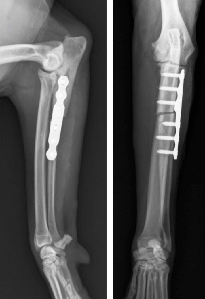
Maybe my sense of perspective is guided by the patients that I see, but I have seen far too many bone on bone elbows the past couple weeks. Evaluation of radiographs or CT images has shown severe osteoarthritis, with osteophytes (bone spurs) and hardened (sclerotic) bone. Looking in the joint with an arthroscope has revealed fragmented coronoid processes, with complete or partial loss of articular cartilage on the weight bearing surface of the ulna and humerus (parts of the elbow joint). Contrary to what the popular press says,there is no osteoarthritis cure! When it gets to this point, the best we can offer are procedures to reduce the weight bearing on these damaged joints, such as shaving down the area with a burr, altering the forces placed on the joint by shifting the weight bearing through corrective osteotomies (cutting a bone and shifting its position to shift the path of weight bearing forces), or performing a partial or complete joint replacement, along with aggressive medical treatment. Even with all of this, the function of affected dogs is fair to poor.
So what is elbow dysplasia? There are 4 main conditions that, collectively, are commonly referred to as elbow dysplasia. Fragmented medial coronoid process (FCP) is the most common condition and is a fragment of the weight bearing surface of the elbow that may be detected between 6-8 months of age. Ununited anconeal process (another part of the ulna in the elbow) is another condition that occurs between 5-6 months of age. The anconeal process is a beak-like projection that is on the notch of the ulna that forms to stabilize the ulna and humerus. Osteochondritis dissecans (OCD) occurs on the medial portion of the humeral condyle as a result of abnormal transition of cartilage to bone as part of the normal growth of joints and bones. Finally, elbow incongruity occurs in some dogs.


So we know the conditions associated with elbow dysplasia, and we know that left unchecked, there will be severe end-stage joint damage. So why isn’t it treated earlier before end stage damage occurs? The problem is not suspecting and recognizing elbow dysplasia at an early age, when fragment removal and corrective osteotomies may slow the progression of arthritis. Notice I said may. Despite optimal treatment at an early age prior to joint deterioration, arthritis will progress. But the hope is that early treatment with continued medical management will result in good function during the life of the dog.
Early recognition is difficult. Most cases have both elbows affected. Visual clinical gait evaluation looks for asymmetry in gait at a walk or trot. Unfortunately, if they are symmetrically bad in terms of reduced weight bearing, we do not see any asymmetry. Furthermore, the degree of forelimb lameness is generally quite severe by the time we can see an asymmetric gait. We have a force platform that allows exact determination of forces placed on each limb. It still amazes me after years of gait evaluation how severe the lameness must be before we see it, with often a 20-30% difference in forces needed before seeing a lameness. Take home message? ANY visual lameness of a forelimb is severe and should be worked up and treated at that time rather than ignoring the problem.
What about radiographs? Sometimes it is possible to see blunting of the coronoid process or even a fragment at an early age. But most of the time, we see arthritic changes on the radiograph, such as osteophytes, or bone spurs, on the anconeal process, head of the radius, and ulna. Increased density (sclerosis) of the trochlear notch of the ulna may also be noted. By the time you can see osteoarthritis on a radiograph, there is irreversible damage to the articular cartilage.

Recently, an article entitled “Epidemiology and clinical management of elbow joint disease in dogs under primary veterinary care in the UK” by O’Neill, Brodbelt, Hodge, Church and Meeson was published in Canine Genetics and Epidemiology. This is an open access journal and the complete article is available for free. This study evaluated canine elbow disease in primary care (not specialty) practices. From 455,069 dogs under veterinary care during a 1 year period, 616 cases of elbow disorders were noted (for comparison, this is a similar prevalence as cruciate ligament disease in the UK assessed by similar study techniques). In my opinion, this number is likely quite low because it is difficult to assess elbow disease without specifically looking for it. All too often, elbow disease is not discovered until there is lameness or pain and the dog is brought to a veterinarian. This is suggested by this study where there were many cases of elbow arthritis diagnosed in middle to late ages of dogs. In fact, the authors indicated that the presenting elbow problems were osteoarthritis in 76%, elbow dysplasia in 31%, and traumatic elbow conditions in 6.7% of dogs.
As noted above, elbow dysplasia occurs in dogs less than one year of age and nearly always leads to arthritis, which may go undetected until later years. Is it possible to have arthritis of the elbow without elbow dysplasia? Sure, it’s possible, but the high numbers reported in the this study seem somewhat unlikely; in my opinion, it is likely that a high number of these cases were secondary to elbow dysplasia, especially fragmented coronoid process, and the authors state that “these later stage presentations are most likely secondary to pre-existing elbow dysplasia but could also result from some forms of primary osteoarthritis”, and “osteoarthritis development in absence of a primary alternative joint disease, so called primary osteoarthritis, is thought to be rare.”
Five breeds showed increased odds of elbow joint disease: Rottweilers, Labrador Retrievers, German Shepherd Dogs, Golden Retrievers and English Springer Spaniels. Ironically, we saw 2 Labrador Retrievers, a Golden Retriever, and a Rottweiler during the past two weeks with end stage elbow arthritis secondary to fragmented medial coronoid processes. Additional risk factors included having an adult bodyweight that was equal or higher than their breed/sex average, advancing age, being male, being neutered, and larger bodyweight. The largest modifiable risk factors are keeping dogs lean and consider delaying the age of neutering.
Some other interesting finding were that the most common signs described by the owners were lameness (76%), difficulty exercising (20%), and pain (14%). The most common findings from veterinary examination were pain (46%), lameness (45%) and reduced range of movement (39%). In a previous study by our own Veterinary Orthopedic Laboratory (link study), we found that ANY loss of elbow range of motion indicated the presence of osteoarthritis and a significant reduction of weight bearing as measured on a force platform. So this is something that can easily be determined in a clinical setting, and ANY loss of elbow range of motion is enough evidence to perform additional diagnostics and initiate aggressive treatment.
We recently reported the results of a study at a meeting of the Veterinary Orthopedic Society describing an approach to the front of the elbow to remove some of the bone spurs that limit joint motion and reduce function. We are excited about a new potential method to manage end stage arthritis and improve function, but there is still severe irreparable damage to the joint.
One very concerning finding in this study from the UK is that of 109 deaths involving euthanasia within the cases of elbow disease, elbow joint disease contributed to the decision to euthanize 41% of the dogs. This means that the elbow disease was so severe in some patients, that it played a major role in the decision to euthanize the dog. Justifiably, the authors concluded that these findings present a clear case for improved breeding programs to reduce the burden of elbow joint disease. Yet, after years of selecting breeding programs, the prevalence of elbow dysplasia remains unacceptably high. So I will go a step further and say that ALL LARGE AND GIANT BREEDS OF DOGS (especially the identified breeds at risk) SHOULD BE SCREENED FOR ELBOW DYSPLASIA BETWEEN 6 AND 10 MONTHS OF AGE. This is important to eliminate affected dogs from the breeding pool and to make sure that young dogs are treated at an early age before debilitating arthritis occurs.
So how is this screening performed? During the final vaccination of dogs between 5 and 8 months of age (especially large and giant breeds), screen for hip dysplasia, elbow dysplasia, and shoulder OCD. Specifically, for elbow dysplasia, palpate both elbow with the dog standing in a square, symmetrical position for any elbow joint effusion or swelling (subtle differences are easier to find with the dog standing and feeling both elbows at the same time). Watch carefully for any conformation problems, especially standing with the toes pointed out. Observe the dog walking and trotting and note ANY gait abnormality. Lay the dog on its side and check for any decreased range of motion or pain at the end of extension or flexion of the joint. With the elbow in mild flexion, place pressure over the medial coronoid process while supinating and pronating the joint (rotating the joint internally and externally) while watching the dog for any signs of pain. If ANY of the above findings are suspect, the next step is imaging. Screening radiographs (especially a flexed lateral view) may find ununited anconeal process and secondary osteoarthritis, and in some instances an abnormal coronoid region. A CT evaluation is more sensitive but is less available and more costly. Diagnostic arthroscopy (a form of surgery using a small camera and instruments to evaluate the joint) is the gold standard, but is more invasive, costly, and is difficult to justify without other findings to support its use.
Call to action
· Have 5 to 9 month old large breed dogs evaluated for orthopedic conditions, especially elbow dysplasia
· Use selective breeding to help reduce the prevalence of elbow dysplasia, especially in breeds at risk
· Keep dogs lean
· Evaluate dogs for elbow conditions throughout their lifetime

Comments