
Elbow Dysplasia
Elbow dysplasia is a common term that results from three different conditions, all of which result in abnormal joint development and secondary osteoarthritis. The three conditions are fragmented medial coronoid process, ununited anconeal process, and osteochondritis dissecans of the medial aspect of the humeral condyle. The elbow is made up of the humerus, radius, and ulna. All three of these bones must come together and fit like a glove. Any shortening or lengthening of any of the three bones results in joint incongruity, which can cause excessive pressure to the other parts of the elbow.
Fragmented Medial Coronoid Process
Fragmented Medial Coronoid process (FMC) is a condition primarily affecting large and giant breeds of dogs, such as Labrador retrievers, Newfoundlands, Bernese Mountain dogs, Rottweilers, etc. Males are affected about twice as frequently as females. There is a heritable component of the disease, so screening of breeding pairs is recommended. The Orthopedic Foundation for Animals (OFA) and the Elbow Working Group in Europe provide assessment of radiographs to screen for elbow conditions. Environmental factors the contribute to the development include excessive weight and rapid growth rate. Clinical signs begin between 5 to 8 months of age, including lameness, swelling of the elbow joint, swinging out of the forelimb to avoid flexing the elbow joint, and short, choppy strides. Approximately 80% of the time, both elbows are affected, therefore asymmetry of gait may not be apparent because both forelimbs are equally affected. Therefore, careful evaluation by your veterinarian at approximately 6 to 8 months of age is critical. In some dogs, a problem is not suspected and lameness may not become apparent until middle age or even later. By this time, there is often end-stage arthritis, with complete loss of cartilage. Therefore, careful evaluation of at-risk breeds by your veterinarian is critical when they are puppies.
Your veterinarian will evaluate your dog's gait to determine if there are any clues suggesting a problem. The elbow joint will be put through a range of motion. Any loss of range of motion of the elbow is associated with osteoarthritis and lameness. Palpation of the elbow joint may reveal swelling or effusion of the elbow joint. Sometimes this is subtle, so palpating both elbows simultaneously in a standing position may allow appreciation of very mild changes. Palpating the area of the medial coronoid process while the elbow joint is rotated inward and outward may demonstrate pain.
Any abnormalities of the gait and orthopedic exams should be followed with imaging, including radiographs and possibly a CT (computerized tomography). Your veterinarian will assess for any abnormalities, including osteoarthritis.

Fragmented Coronoid Process
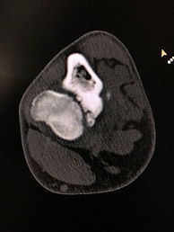
Fragmented Coronoid Process
If a FCP is identified at an early age prior to significant arthritis development, arthroscopic removal of the fragment is generally recommended. If the joint has a "step" incongruity, surgical correction may be performed by doing an osteotomy of one of the bones to allow the joint to become more normal. The osteotomy may be a simple oblique cut of a bone, or involve special implants, such as with a proximal abducting ulnar osteotomy (PAUL).

PAUL procedure
As the condition progresses such that cartilage loss occurs, other treatments may be necessary, including:
-
arthroscopic removal of the fragment with microfracture (micropick) of the exposed bone to allow blood supply and fibrocartilage formation (not articular cartilage, but a type of cartilage to cover the bone)
-
sliding humeral osteotomy (SHO), which involves an osteotomy of the humerus, with shifting of the bone segments to reduce weightbearing on the diseased medial compartment of the elbow
-
canine unicompartmental elbow (CUE), which is a partial elbow replacement
-
total elbow replacement - this procedure carries a high complication rate and careful consideration should be made before embarking on this treatment
Osteochondritis Dissecans (OCD)
of the medial aspect of the humeral condyle
OCD can occur in a number of joints, including the elbow, shoulder, stifle, and hock. It has a genetic basis, as well as environmental factors, including rapid growth rate, obesity, and high loads on joints. The condition occurs when joint cartilage does not turn into bone as part of the normal growth process, resulting in excess cartilage thickness. This area is weak, and as a result, a fragment may break off. In the elbow, the result is pain, lameness, and arthritis.
Ununited Anconeal Process (UAP)
UAP is a condition in which a growth plate fails to close at the appropriate time, and the fragment remains loose, causing pain, lameness, and arthritis. This growth plate normally closes by 5 1/2 months of age. If it remains open at 6 months, it is considered to be a UAP. This condition is most common in German Shepherd dogs and Bassett hounds. If it is found before severe arthritis is present, surgery to correct the joint incongruity (osteotomy of the ulna) and screw fixation of the UAP may be performed. Unfortunately, most cases are undiagnosed until moderate to severe arthritis are present. In this case, surgical removal of the fragment is performed, followed by arthritis management.

Ununited Anconeal Process
Secondary Arthritis
All of the components of elbow dysplasia result in osteoarthritis. Please refer to the section on arthritis for ideas regarding management of arthritis. Even if surgery is performed at an early age, arthritis progresses and requires treatment.

Biceps Insertional Tendinopathy
This is a relatively newly recognized condition. Dogs can be quite lame and painful. Many dogs that are affected play quite hard, with much running and sharp turns. The result is pain and inflammation at the insertion of the biceps brachii muscle on the ulna and radius, just below the front (cranial) part of the elbow joint. Dogs are quite sensitive on palpation of this area. The biceps muscle is located on the craniomedial (front inside) part of the humerus, and it is followed distally (down the leg) to its insertion. Some extension of the elbow joint makes the muscle and its tendon more distinct. The area of the tendon insertion is distinct from the area of the medial coronoid process, which is painful when there is a fragmented medial coronoid process. Treatment involves rest and anti-inflammatory medication. Also, treatment with extracorporeal shockwave treatment for 1 to 4 treatments seems to help patients recover more quickly. Recurrence is possible.